
A Guide to Clinical Differential Diagnosis of Oral Mucosal Lesions
About This Event
This self-instructional course provides an overview of common oral mucosal entities, including their presentation, management and diagnosis. The primary goal is to teach dental professionals the process of clinical differential diagnosis of diseases and lesions of the oral mucosa. The course includes an interactive and downloadable decision tree to assist diagnosis, and references to clinical image atlases (The Oral Pathology Image Database). Content is designed to be used with the atlas; lesions with available clinical images are designated with an asterisk. Course includes authorship and conflict-of-interest disclosures, recommended additional resources, and is approved as a PACE program.
Learning Objectives
- Classify oral lesions into surface lesions and soft tissue enlargements using a decision tree (flowchart).
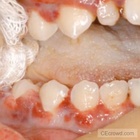
- Describe the clinical features characteristic of each class of oral mucosal lesions in the decision tree, including white surface lesions, generalized pigmented surface lesions, localized pigmented surface lesions, vesicular-ulcerated-erythematous surface lesions, reactive soft tissue enlargements, benign tumors, malignant neoplasms, and cysts of oral mucosa.
- Describe the characteristic or unique clinical features of the most common and/or important diseases of the oral mucosa.
- Perform a step-by-step clinical differential diagnosis, using the decision tree, for patients with oral mucosal lesions.
Additional Event Information
Target Audience
Dental Assistant Students, Dental Assistants, Dental Hygiene Students, Dental Hygienists, Dental Students, Dentists
Event Format
Virtual
CE Credits
4*